


Diagnostic Image Department
This is a specialized department that mostly deals with producing images of the anatomy and use these images to aid in the diagnosis. Our department make use of many specialized machinery in the medical field, especially in the use of X-ray, Ultrasounds, Electromagnetic wave etc.
Aek Udon International hospital is well equipped with the most advanced medical imaging facilities and equipment, along with a team of professional radiologists who is able to carry diagnosis in effective and timely manner.
Diagnostic radiology equipment consists of
1. Digital Radiography machine is capable of presenting and storing detailed digital images with precision and accuracy as well as storing the amount of radiations that the skin of the patients receive, together with images, and then put the information on the picture-archiving and communication system(PACC). With the Autonomic Exposure Control technology, the digital radiography is able to consistently control the amount of radiation.
Aek Udon is the only hospital in the Northeast that has been appointed by the Government of Australia and New Zealand to conduct the immigration medical examination for long term visa applicants.
Fluoroscopy is an imaging technique that uses X-rays to obtain real-time moving images of the interior of an object.
2. Fluoroscopy is an imaging modality that uses x-rays to allow real-time visualization of body structures. This allows for dynamic assessment of anatomy and function. High density contrast agents may be introduced into the patient to allow for greater differentiation between structures and greatly helps with the diagnosis.
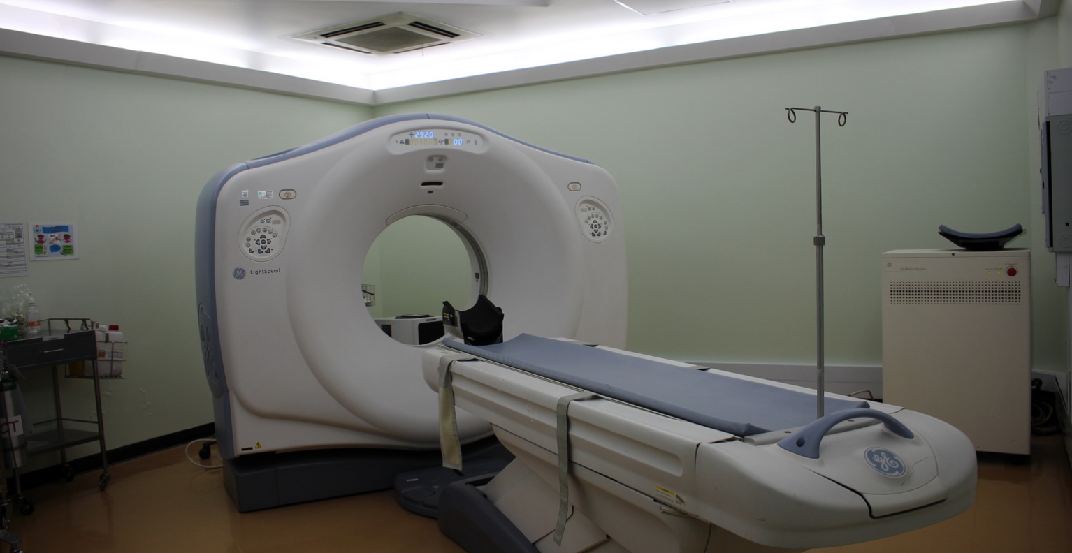
3. The computerized tomography(CT) scan combines a series of X-ray images taken from different angles around your body and uses computer processing to create cross-sectional images (slices) as well as 3D images of the bones, blood vessels and soft tissues inside the body.
4. An ultrasound scan uses high frequency sound waves to examine the internal organs by utilizing the sound absorption properties and the fact that different organs reflect sound waves differently in order to create images
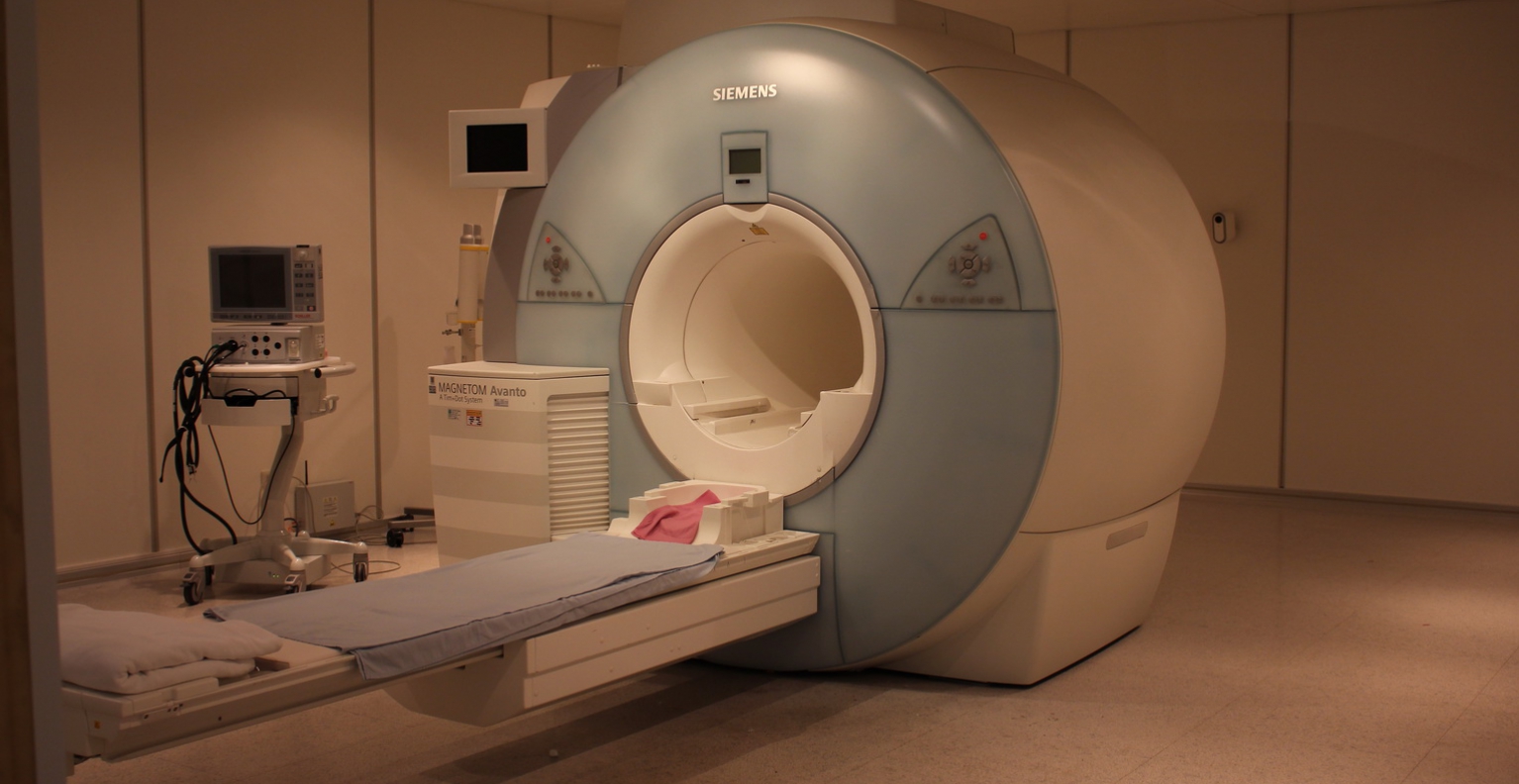
5. Magnetic Resonance Imaging (MRI) machine uses strong magnetic field and up-to-date technology to check and produce detailed cross-sectional images as well as 3D images of the internal organs. The MRI machine is very safe and does no cause radiation accumulation in the body.
6. The Digital Mammography is used specifically for the examination of breasts and the storage of detailed digital photography with precision and accuracy. The Digital Mammography is able to detect early stages of breast cancer as well as metastatic stages of breast cancer.
7. Bone mineral density scan uses low power X-rays to estimate the density and thickness of the bone with great accuracy. The bone mineral density scan has been set as a standard test for osteoporosis by the World Health Organization.
8. The Digital Dental X-ray scanner is capable of examining teeth as well as displaying and storing detailed digital images with precision and accuracy. Hence, this can assist in the diagnosis and treatment planning as well as helping with dental implant, root canal treatment and oral surgery.